Pages

Thursday, 11 June 2015

The Kids Aren’t Alright – Part II


One of the many misconceptions around endometriosis is that it is a disease solely afflicting adult women in their mid-thirties. This belief was prevalent for many, many years and still lingers today. However, whilst it may be true that most women with endo are usually diagnosed in their twenties or thirties, the truth is the symptoms of endo appear far earlier and, through dismissal of symptoms leading to diagnostic delay, the true age at which endometriosis presents is overlooked.

Fortunately today there seems to be a drive to increase study and awareness about endometriosis in young women. Today I am going to discuss one such study from the US, a free, full text version of the article you can find here.

This study included 25 cases of girls under 21 years old undergoing laparoscopy for pelvic pain who had no previous diagnosis of endometriosis either from laparoscopy or radiological methods (ultrasound, MRI etc). Information was collected from all patients before and after surgery to see what the characteristics of endometriosis in these patients could tell us about adolescent endo (albeit in a small cohort, but you’ve got to start somewhere).

The average age of the patients in this group was 17.2 years old. Interestingly 14 out of the 25 (56%) reported a family history of endometriosis. This is far higher than what you would expect if it was a group of girls selected at random from the general population. What this means is that risk of developing endometriosis at a young age is significantly increased by a family history of endo, a fact that doctors and women with endo who have daughters need to be very aware of. The results of this study do seem to suggest that parents with endo are more than capable of advocating their case though, as 44% of referrals came from the patient’s mother.

In terms of the symptoms the girls experienced, the most common gynaecological symptoms were the ones most typically associated with endo, such as dysmenorrhoea (excessively painful periods) in 64% and abnormal/irregular bleeding in 60%. This is an issue of particular importance as these symptoms can lead to days missed from school every month, potentially damaging a young girls prospects in adulthood. Only 4 out of the 25 patients reported dyspareunia (painful sex), but being as only 8 out of the 25 reported being sexually active, dyspareunia is a poor measure of endo risk in this population. 

Of the gastrointestinal symptoms nausea was the most common, being present in nearly half (44%) of all patients. Between a quarter and fifth of all patients experienced some other gastro symptoms, such as constipation or diarrhoea. Fatigue, an often unrecognised symptom of endo, was also present in around a quarter of all patients and seems to get more common with age.

A very high degree of variation was reported in the time it took from the initial visit to a physician to diagnosis. The range in this cohort varied from 1 month to 9 years, with the delay between onset of symptoms and diagnosis being 2 years on average. This just goes to show how important it is for doctors to be well educated in recognising the signs of endometriosis in adolescent girls and adult women.

The authors of this study mentioned another survey of over 4000 women diagnosed with endo. Two thirds of these women said their symptoms appeared during adolescence and that their symptoms were far less likely to be taken seriously when they were young that when they were adults.

After the 25 patients had received surgery it was discovered that 17 had stage I disease, 5 had stage II, 3 had stage II and none had stage IV. These are similar findings to a study published just a few weeks earlier which included 55 girls aged under 19 who were found to have endo. It is also unsurprising to find that no cases of stage IV endo as previous studies also report a very low rate of ovarian endometriotic cysts in adolescents. However, severe stage endometriosis in young women is not unheard of, particularly after the age of 17, and therefore should not be dismissed. An observation made by the authors was that the appearance of endometriosis in adolescents can be different to that of adult women. For example, they noted that the predominant lesion types were subtle atypical lesions like clear, white and red, whereas in adult women surgeons would be more likely to see the darker, blueish-black lesions. This is an important factor that needs to be taken into consideration by surgeons as the subtler forms of endo can be easily missed.

After a 1 year follow up 80% of the patients had improved or resolved pain, however this was a relatively short follow up period and, as different patients received different post-operative treatment, it’s hard to say how this affected the resolution of symptoms.

Nevertheless, this article raises several important issues surrounding endometriosis in young women. In particular how it can present in a different manner, both symptomatically and physically, to endo in adult women and how better characterisation of adolescent endometriosis can lead to quicker diagnosis, better treatment outcomes and an overall lessening of the burden of endometriosis on women and society.

Tuesday, 2 June 2015

A Seminal Piece of Work


Remember when you are at school and there would always be rumours and stories going around the playground, like eating popping candy and drinking cola would make your stomach explode. Well it seems like the media’s attempts at reporting scientific news has become the new school playground.

I say this for several reasons, but particularly because of reports I’ve seen online recently concerning a study into the effect of seminal fluid on endometriosis. Seminal fluid is the major component of semen and, if these reports are to be believed then “It is now understood that exposure of the endometrium, which is the inner lining of the uterus, to seminal fluid may contribute to the progression of the endometriosis in women.” That is a direct quote from one web site attempting to give an account of some new research that was published recently and, in typical fashion, they have misquoted the original research, probably didn’t read the original article and relied solely on vox pops and sensationalism.

Let’s go back to the original research then, which was published here and a press release from the university it was conducted at was written here. For some reason the journal in which the research was published is probably the only journal my university doesn’t have a subscription to, so I can’t read the full article. Therefore the comments I’ll make here are based on the abstract for the article.

To start then these researchers are interested in transforming growth factor beta (TGFβ), a chemical messenger in your body that is involved in cell growth and death. I use the word TGFβ as a singular, but it actually refers to a whole family of messengers comprising over 30 members with a variety of different functions. To be more specific then, these researchers found that a deficiency of TGFβ-1 in mice impaired their development of experimentally induced endometriosis, implicating TGFβ-1 as important for the establishment and growth of endo.  Human seminal fluid has a high concentration of TGFβ, therefore the researchers postulated that exposure so seminal fluid may influence the growth of endometriosis.
To test this they took samples of human endometrium, exposed some of the samples to seminal fluid and some to a normal growth solution. They then surgically implanted the endometrial samples in mice and after two weeks noted that the endometrial tissue that had been exposed to seminal fluid had just over a fourfold increase in volume. On the face of it then it would seem that exposure to seminal fluid does increase the growth of endometriotic lesions. However there are several assumptions made in this study that could impact the relevance of the results.

  •  It assumes the normal endometrium is the same as endometriosis. Granted the endometrium and endometriosis do look similar, however in recent years it has been discovered that there are hundreds of differences between them. Therefore the way an experimentally induced endometriotic lesion reacts to a stimulus could be very different from the way a human lesion reacts. 
  •  I couldn’t find out whether or not the researchers used the endometrium from a woman with or without endometriosis, but this is also a key point. The normal endometrium from women with and without endo differs in a number of aspects. Of particular relevance is the fact that the endometrium of women with endo has a higher level of TGFβ-1 than women without endo. This could mean that endometrial cells from women with endo are more sensitive to TGFβ (but that depends on whether they have the receptors to recognise TGFβ as well). 
  •  Animal models are a continuing source of contention. Unfortunately though they are the only way in which complex diseases can be studied in a living system, so until something better comes along we’re stuck with them. The first issue is that the mice used were immunodeficient i.e. their immune systems were dampened down so they wouldn’t reject the implanted endometrium). This could mean that the way in which the endometriotic implant grew in the animal was different to how it would grow in a human. Secondly, mice don’t have the same menstrual cycle as humans, in fact they don’t have a menstrual cycle at all, they have an estrous cycle. Estrogen and progesterone can alter the production of TGFβ-1 by the endometrium so the cyclic variation and the effect of hormone therapies needs to be taken into consideration. 

  • Another thing I couldn’t find out is what the dose of seminal fluid they pre-treated the endometrium was before implanting it in the mouse. This is important because we need to know whether it was physiologically relevant i.e. does the dose they used correspond to what would be found in the human body, in particular, how much seminal fluid actually reaches the pelvic cavity after intercourse and could come into contact with endometriosis.

  •  The peritoneum (the layer of tissue that surrounds the reproductive organs) secretes TGFβ-1 itself in women with endometriosis leading to an increase in TGFβ-1 in peritoneal fluid from women with endo. Therefore the amount of TGFβ-1 that seminal fluid contributes may be insignificant compared to the amount already produced around the endometriotic lesions by the peritoneum. 


Overall then it is hard to say whether human endometriosis is ever exposed to seminal fluid or if it can influence the growth of endometriosis in humans, certainly I don’t see any evidence to make me believe exposure to seminal fluid causes endometriosis. For some women with endometriosis sex can lead to pain both during and after, but I would think this is down to pressure and torsion put on the pelvic area that is already sensitised due to of endometriosis, rather than exposure to components of semen.  

Whilst there are limitations to this research, it does give us some interesting points to think about. In particular the effect of TGFβ on the normal endometrium, how this differs between women with and without endometriosis and how this could affect fertility. For example, TGFβs are suspected to be involved in the early development of the embryo and their production changes throughout the menstrual cycle. Finding out if these changes are altered in women with and without endo and if this effects the growth and receptivity of the endometrium is definitely worth investigating.    

Friday, 20 March 2015

Endometriosis Awareness Month - Part III


Endometriosis is a puzzling disease, there are still many unanswered question surrounding its origin, who gets it and why. In this way it makes studying endometriosis like exploring the world in the 17th century, there is a sense of undiscovered territory to explore, but rather than large wooden ships and subjugating natives, we have theories and hypotheses to test with science. Some of those theories prove worth exploring, some have a little, a lot, or no evidence to support them. Creating new theories sometimes seems like muddying the water and complicated an already complicated subject, but it is only by coming up with and testing new ideas, we get closer to the truth (however, all ideas must be testable and all tests must be fair, objective and subject to scrutiny by peer review, something pseudoscience always fails at).

With that in mind I’m going to be doing a bit of theorising myself in this week’s blog post. A big part of my research is examining the role that inflammation plays in endometriosis, specifically what controls inflammation in endometriotic and normal endometrial tissue. Part of this research involves looking into the initial causes of inflammation, and one thing in particular has caught my attention recently and that is bacteria.

Some types of bacteria are essential for our health, without them we would become very ill indeed and several of our organs have their own population of micro-organisms that help them function correctly. The gut and the vagina are two good examples, the gut in particular contains between 500 and 1000 different species of bacteria, numbering in the trillions of individual micro-organisms. Interestingly, the harmless bacteria that naturally populate the vagina actually help prevent infections by more harmful bacterial types. But sometimes disruption of the natural order of our internal microflora can lead to harmful effects.

Now I’m not falling into the trap of confusing endometriosis with endometritis and I’m certainly not suggesting that endometriosis is an infection or caused by an infection. Rather bacteria may play a hitherto under-recognised role in inflammation and the generation of painful symptoms in endometriosis.

How might bacteria contribute to endometriosis? To clarify I’m talking about a specific type of bacteria, called Gram-negative bacteria, which is a group of bacteria including E.coli, one of the better known species of bacteria. Why these organisms are important is because of something they secrete, namely lipopolysaccharides (LPS). LPS are essentially toxins the bacteria produce that are responsible for making you ill, but they are also powerful stimulants for inflammation and this is the key point of interest.

To start with we need to know which bacteria are normally present in the uterus and how this differs in women with endometriosis. A study published in 2014 investigated this very phenomena and found that several bacteria types, especially E.coli, were increased in the endometrium of women with endometriosis (see the figure below)


The solid white boxes represent the levels of micro-organisms found in the endometrium of women without endo, the dashed boxes represent the levels in women with endo.Figure source: http://humrep.oxfordjournals.org/content/29/11/2446.long

The solid white boxes represent the levels of micro-organisms found in the endometrium of women without endo, the dashed boxes represent the levels in women with endo.

What we can see from this is that there are several types of micro-organism found at increased levels in the endometrium of women with endometriosis, in particular a large increase in E.coli. Following on from this further studies found that LPS was found at a much higher concentration in the menstrual fluid and peritoneal fluid of women with endo as you can see if you click this link and look at figures C and D (the white bars represent the levels of LPS in women without endo, the black bars are women with endo). 

The increase in LPS in the menstrual fluid can be attributed to the increase in E.coli in the endometrium, but what about the peritoneal fluid? This can be explained by retrograde menstruation, where menstrual fluid is passed upwards through the fallopian tubes and out into the peritoneal cavity. Retrograde menstruation is a natural event in the female body during menstruation, occurring in as much as 90% of women and it was long thought that this was the root cause of endo, with endometrial cells shed during a period being passed into the peritoneal cavity where they implant and grow. This theory has since fell from favour somewhat as more evidence accumulates against it being the cause of endo, but there may be a role for retrograde menstruation in endometriosis, just perhaps not what was previously thought.

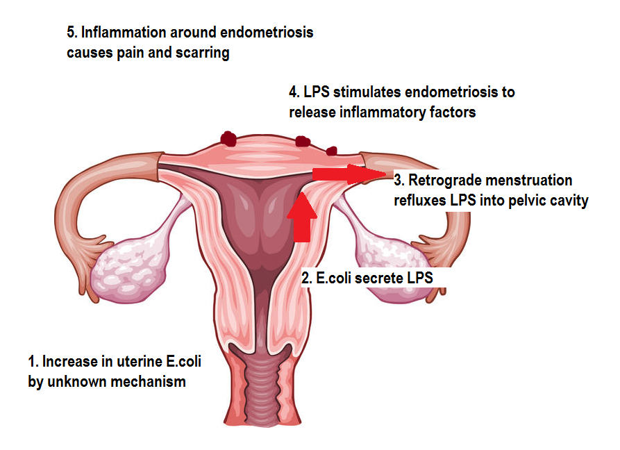
It seems plausible then that increased levels of E.coli in the endometrium lead to higher concentrations of LPS in the menstrual blood, which can get refluxed into the peritoneal cavity by retrograde menstruation, increasing the concentration of LPS in the peritoneal fluid, which can be in close/direct contact with endometriotic lesions. The next step is to assess what can happen when
LPS interacts with endometriosis.

Some studies have looked into this and found that LPS can stimulate endometriotic cells to release factors, like prostaglandin E2 (PGE2) and tumour necrosis factor (TNFα)  which promote inflammation and stimulate pain signals (PGE2 has been implicated in a number of important facets for endometriosis survival including cell growth, inhibition cell destruction and hormone production). This would lead to a heightened inflammatory state around endometriotic lesions and in the normal endometrium, possibly contributing to symptoms frequently associated with endometriosis, such as chronic pelvic pain and excessively painful periods. The whole process can be summed up on the diagram below.


Original image from https://edc2.healthtap.com/ht-staging/user_answer/reference_image/7081/large/Uterus.jpeg?1386669522

The final question, which we don’t have an answer to yet, is why E.coli and other micro-organisms are increased in the endometrium of women with endo.  It may be due to alterations of vaginal acidity leading to easier colonisation of the uterus by bacteria, or dysfunction of the immune cells in the uterus of women with endo resulting in them not being able to keep micro-organisms in check? Also, what effect would restoring the normal balance of uterine micro-organisms have and how can this be achieved? There are a lot of issues that need to be addressed here, but hopefully this will be an area of investigation that yields positive results in terms of relief from painful symptoms.

Monday, 9 March 2015

Endometriosis Awareness Month - Part II


Each year endometriosis awareness month seems to get bigger and bigger. I would highly recommend checking out the hashtag #endometriosis if you’re on Twitter to keep up with all the news and events from around the world.

For this week’s blog post I’m going to be putting some ideas out there about the origin of endometriosis. Not the way in which the disease originates in the body (there could be a whole book written on that subject), but where and when endometriosis actually came from in the history of humanity.

Some people tend to think of endometriosis as ‘modern’ condition, however there are several great resources examining the history of endometriosis, for example Dr Nezhat’s paper and Dr Redwine’s book, which suggest that endometriosis has been known about for thousands of years under different names and guises. But this week I’m going even further back, to the beginning.

With that in mind let’s start at the beginning, which is always a good place to start. In order to find the origin of endometriosis I started by searching for which animals are known to develop endo spontaneously (there are numerous animals that can be induced to have ‘experimental endometriosis’ but I’m only interested in animals in which endo has been found to occur naturally). Having a thorough search through the literature I discovered endo has been documented in the following animals (see the end of the blogpost for the references).

Human (obviously)          Rhesus Macaque
Gorilla                              Cynomolgus monkey
Olive Baboon                   Taiwan Macaque
Yellow Baboon                 Pigtail Macaque
Hamandryas Baboon        Debrazza’s monkey
Guinea Baboon                African Green monkey
Dog Face Baboon            Gray-cheeked mangabey
Mandrill                           White-tufted-ear marmoset

One interesting thing you may notice straight away is that all the species noted above are primates. Endometriosis has never been documented as occurring in any non-primate species (there is a condition affecting livestock called endometriosis, but I won’t be counting that as it refers to degeneration of the endometrium, not the same thing as the real endometriosis). This tells us there is something these animals have in common, not shared by other animals that allows them to develop endometriosis. Given that the majority of these cases of endometriosis  in primates have been discovered by accident or when performing autopsies on the animals, it is highly likely that there are other primate species in which endo is yet to be discovered (chimpanzee’s, our closest relatives, are conspicuously absent from the list, for example).

What can we do then with the above information? To answer that lets look at how all those species listed above relate to one another. We know from 150 years of evolutionary biology that all living creatures on earth are related to one another in a fantastically complex tree of life of which we are only a tiny branch.

http://www.utexas.edu/features/graphics/2008/tree/tree3.jpg


(for a more detailed look at our place in the tree of life, see here http://www.zo.utexas.edu/faculty/antisense/tree.pdf)

Zooming in on the part of that tree that contains the primates we can see our relation to those animals closest to us.

Click to enlarge


Believe it or not, this is a simplified version of the primate family tree, there are numerous species that I’ve left out because they’re extinct and I’ve omitted several species and grouped together others to keep it as simple as I can whilst retaining the important information. Now if we connect all the species in which endo has been found, we can see something very interesting.

Click to enlarge


The red lines connect all the species in which endometriosis has been documented thus far, so now we see the natural history of endometriosis. The arrow near the bottom shows the point at which, from the evidence we have, it is plausible to suggest endometriosis originated. Whatever long extinct animal lived at this point, it passed on the predisposition to endometriosis to its relatives. What that predisposition was exactly remains to be clarified. It must be a genetic predisposition, but knowing exactly what gene or genes are responsible we cannot say. Mostly because there is little agreement as to what genes are associated with endo in humans, let alone across the boundary of species. Whatever it was it changed something, perhaps the way in which the reproductive system developed, perhaps the way in which the reproductive organs function, at the moment we can’t say.

If we suppose that endometriosis did originate at this point, we can now theorise as to when endometriosis arose by finding when the species it originated in lived. Looking at where all the red lines converge we can see it is at the point in time when the group of primates containing the marmosets (called the New World Primates) split from the group of primates containing humans, chimps, macaques etc (called the Old World Primates). The common relative of these groups is thought to have lived in north Africa near Egypt 1,2, as this is where the highest proportion of such fossils are found. Analysis of the age of these fossils put them at around 33-35 million years old 2. However, using genetic analysis to date species separation based on how closely related they are puts the date at around 43 million years ago 3. From this evidence we can tentatively theorise that endometriosis could be tens of millions of years old; older than human history records, older than humanity itself.

We tend to think of endometriosis in terms of human history and why shouldn’t we? Endometriosis affects us in the here and now, but if we consider our past it may give us hints to the answers we’ve been looking for. For example, if we know what animals can develop endo and which ones cannot, then perhaps we should find out why that is. What is it about these animals that make them different? By answering this question we could finally understand what predisposes an individual to endo and maybe even how to stop it.


11.  Fleagle J. Primate adaptation and evolution 2nd Edition (Academic Press, San Diego, 1999)
22.  Kappelman J, Simons E, Swisher C. New age determinations for the Eocene-Oligocene boundary sediments in the Fayum depression, northern Egypt. J Geol, 100, 647-668 (1992)
33.  Steiper ME, Young NM. Primate molecular divergence dates. Mol Phylogenet Evol, 41(2), 384-394 (2006).

References for cases of primate endometriosis

Gorilla: Doré M, Lagacé A. Spontaneous External Endometriosis in a Gorilla (Gorilla gorilla). Can Vet J, 26(11), 347-349 (1985)
Olive baboon and Yellow baboon: D'Hooghe TM, Bambra CS, De Jonge I, Lauweryns JM, Raeymaekers BM, Koninckx PR. The effect of pregnancy on endometriosis in baboons (Papio anubis, Papio cynocephalus). Archives of gynecology and obstetrics, 261(1), 15-19 (1997).
D'Hooghe TM, Bambra CS, Raeymaekers BM, Koninckx PR. Serial laparoscopies over 30 months show that endometriosis in captive baboons (Papio anubis, Papio cynocephalus) is a progressive disease. Fertility and sterility, 65(3), 645-649 (1996)
Hamadryas baboon: Shalev M, Ciurea D, Deligdisch L. Endometriosis and stromal tumor in a baboon (Papio hamadryas). Laboratory animal science, 42(2), 204-208 (1992).
Guinea baboon: Dallwig RK, Langan JN, Hatch DA, Terio KA, Demitros C. Bilateral hydronephrosis secondary to endometriosis managed by endoscopic ureteral stent placement in a captive Guinea baboon (Papio papio). J Zoo Wildl Med, 42(4), 747-750 (2011).
Dog face baboon: Folse DS, Stout LC. Endometriosis in a baboon (Papio doguera). Laboratory animal science, 28(2), 217-219 (1978)
Rhesus macaque: Zondervan KT, Weeks DE, Colman R et al. Familial aggregation of endometriosis in a large pedigree of rhesus macaques. Human reproduction (Oxford, England), 19(2), 448-455 (2004)
Crab eating macaque: Fanton JW, Hubbard GB. Spontaneous endometriosis in a cynomolgus monkey (Macaca fascicularis). Laboratory animal science, 33(6), 597-599 (1983)
Formosan macaque: Chin S. Endometriosis and pyometra in a Taiwan rhesus (Macaca cyclopis). J Chin Soc Vet Sci, 20, 58-64 (1994)
Pigtail macaque: DiGiacomo RF, Hooks JJ, Sulima MP, Gibbs CJ, Jr., Gajdusek DC. Pelvic endometriosis and simian foamy virus infection in a pigtailed macaque. Journal of the American Veterinary Medical Association, 171(9), 859-861 (1977)
De Brazza’s monkey: Binhazim AA, Tarara RP, Suleman MA. Spontaneous external endometriosis in a De Brazza's monkey. J Comp Pathol, 101(4), 471-474 (1989)
African green monkey: Cary C, Peter G, Schiffer S. A case report and review of endometriosis in nonhuman primates. Laboratory animal science, 32, 426 (1982)
Magabey: Schmidt R, Hartfiel D. Endometriosis in a Gray-Cheeked Mangaby [Cercocebus albigena]. J Zoo An Med, 9(2), 42-45 (1978).
Mandrill: Pirarat N, Kesdangsakolwut S, Chotiapisitkul S, Assarasakorn S. Spontaneous diabetes mellitus in captive Mandrillus sphinx monkeys: a case report. J Med Primatol, 37(3), 162-165 (2008).
Nakamura S, Ochiai K, Ochi A, Ito M, Kamiya T, Yamamoto H. Spontaneous Endometriosis in a Mandrill (Mandrillus sphinx). J Comp Pathol).
Marmoset: Spontaneous pathology of the common marmoset (Callithrix jacchus) and tamarins (Saguinus oedipus, Saguinus mystax). J Med Primatol, 38(5), 347-359 (2009).