Pages

Friday 20 March 2015

Endometriosis Awareness Month - Part III


Endometriosis is a puzzling disease, there are still many unanswered question surrounding its origin, who gets it and why. In this way it makes studying endometriosis like exploring the world in the 17th century, there is a sense of undiscovered territory to explore, but rather than large wooden ships and subjugating natives, we have theories and hypotheses to test with science. Some of those theories prove worth exploring, some have a little, a lot, or no evidence to support them. Creating new theories sometimes seems like muddying the water and complicated an already complicated subject, but it is only by coming up with and testing new ideas, we get closer to the truth (however, all ideas must be testable and all tests must be fair, objective and subject to scrutiny by peer review, something pseudoscience always fails at).

With that in mind I’m going to be doing a bit of theorising myself in this week’s blog post. A big part of my research is examining the role that inflammation plays in endometriosis, specifically what controls inflammation in endometriotic and normal endometrial tissue. Part of this research involves looking into the initial causes of inflammation, and one thing in particular has caught my attention recently and that is bacteria.

Some types of bacteria are essential for our health, without them we would become very ill indeed and several of our organs have their own population of micro-organisms that help them function correctly. The gut and the vagina are two good examples, the gut in particular contains between 500 and 1000 different species of bacteria, numbering in the trillions of individual micro-organisms. Interestingly, the harmless bacteria that naturally populate the vagina actually help prevent infections by more harmful bacterial types. But sometimes disruption of the natural order of our internal microflora can lead to harmful effects.

Now I’m not falling into the trap of confusing endometriosis with endometritis and I’m certainly not suggesting that endometriosis is an infection or caused by an infection. Rather bacteria may play a hitherto under-recognised role in inflammation and the generation of painful symptoms in endometriosis.

How might bacteria contribute to endometriosis? To clarify I’m talking about a specific type of bacteria, called Gram-negative bacteria, which is a group of bacteria including E.coli, one of the better known species of bacteria. Why these organisms are important is because of something they secrete, namely lipopolysaccharides (LPS). LPS are essentially toxins the bacteria produce that are responsible for making you ill, but they are also powerful stimulants for inflammation and this is the key point of interest.

To start with we need to know which bacteria are normally present in the uterus and how this differs in women with endometriosis. A study published in 2014 investigated this very phenomena and found that several bacteria types, especially E.coli, were increased in the endometrium of women with endometriosis (see the figure below)


The solid white boxes represent the levels of micro-organisms found in the endometrium of women without endo, the dashed boxes represent the levels in women with endo.Figure source: http://humrep.oxfordjournals.org/content/29/11/2446.long

The solid white boxes represent the levels of micro-organisms found in the endometrium of women without endo, the dashed boxes represent the levels in women with endo.

What we can see from this is that there are several types of micro-organism found at increased levels in the endometrium of women with endometriosis, in particular a large increase in E.coli. Following on from this further studies found that LPS was found at a much higher concentration in the menstrual fluid and peritoneal fluid of women with endo as you can see if you click this link and look at figures C and D (the white bars represent the levels of LPS in women without endo, the black bars are women with endo). 

The increase in LPS in the menstrual fluid can be attributed to the increase in E.coli in the endometrium, but what about the peritoneal fluid? This can be explained by retrograde menstruation, where menstrual fluid is passed upwards through the fallopian tubes and out into the peritoneal cavity. Retrograde menstruation is a natural event in the female body during menstruation, occurring in as much as 90% of women and it was long thought that this was the root cause of endo, with endometrial cells shed during a period being passed into the peritoneal cavity where they implant and grow. This theory has since fell from favour somewhat as more evidence accumulates against it being the cause of endo, but there may be a role for retrograde menstruation in endometriosis, just perhaps not what was previously thought.

It seems plausible then that increased levels of E.coli in the endometrium lead to higher concentrations of LPS in the menstrual blood, which can get refluxed into the peritoneal cavity by retrograde menstruation, increasing the concentration of LPS in the peritoneal fluid, which can be in close/direct contact with endometriotic lesions. The next step is to assess what can happen when
LPS interacts with endometriosis.

Some studies have looked into this and found that LPS can stimulate endometriotic cells to release factors, like prostaglandin E2 (PGE2) and tumour necrosis factor (TNFα)  which promote inflammation and stimulate pain signals (PGE2 has been implicated in a number of important facets for endometriosis survival including cell growth, inhibition cell destruction and hormone production). This would lead to a heightened inflammatory state around endometriotic lesions and in the normal endometrium, possibly contributing to symptoms frequently associated with endometriosis, such as chronic pelvic pain and excessively painful periods. The whole process can be summed up on the diagram below.


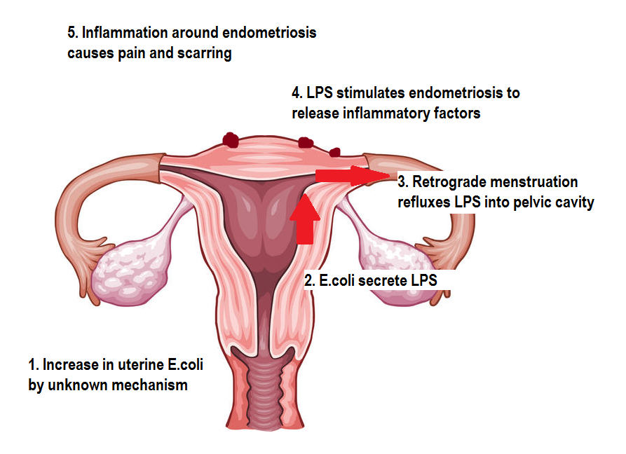
Original image from https://edc2.healthtap.com/ht-staging/user_answer/reference_image/7081/large/Uterus.jpeg?1386669522

The final question, which we don’t have an answer to yet, is why E.coli and other micro-organisms are increased in the endometrium of women with endo.  It may be due to alterations of vaginal acidity leading to easier colonisation of the uterus by bacteria, or dysfunction of the immune cells in the uterus of women with endo resulting in them not being able to keep micro-organisms in check? Also, what effect would restoring the normal balance of uterine micro-organisms have and how can this be achieved? There are a lot of issues that need to be addressed here, but hopefully this will be an area of investigation that yields positive results in terms of relief from painful symptoms.

Monday 9 March 2015

Endometriosis Awareness Month - Part II


Each year endometriosis awareness month seems to get bigger and bigger. I would highly recommend checking out the hashtag #endometriosis if you’re on Twitter to keep up with all the news and events from around the world.

For this week’s blog post I’m going to be putting some ideas out there about the origin of endometriosis. Not the way in which the disease originates in the body (there could be a whole book written on that subject), but where and when endometriosis actually came from in the history of humanity.

Some people tend to think of endometriosis as ‘modern’ condition, however there are several great resources examining the history of endometriosis, for example Dr Nezhat’s paper and Dr Redwine’s book, which suggest that endometriosis has been known about for thousands of years under different names and guises. But this week I’m going even further back, to the beginning.

With that in mind let’s start at the beginning, which is always a good place to start. In order to find the origin of endometriosis I started by searching for which animals are known to develop endo spontaneously (there are numerous animals that can be induced to have ‘experimental endometriosis’ but I’m only interested in animals in which endo has been found to occur naturally). Having a thorough search through the literature I discovered endo has been documented in the following animals (see the end of the blogpost for the references).

Human (obviously)          Rhesus Macaque
Gorilla                              Cynomolgus monkey
Olive Baboon                   Taiwan Macaque
Yellow Baboon                 Pigtail Macaque
Hamandryas Baboon        Debrazza’s monkey
Guinea Baboon                African Green monkey
Dog Face Baboon            Gray-cheeked mangabey
Mandrill                           White-tufted-ear marmoset

One interesting thing you may notice straight away is that all the species noted above are primates. Endometriosis has never been documented as occurring in any non-primate species (there is a condition affecting livestock called endometriosis, but I won’t be counting that as it refers to degeneration of the endometrium, not the same thing as the real endometriosis). This tells us there is something these animals have in common, not shared by other animals that allows them to develop endometriosis. Given that the majority of these cases of endometriosis  in primates have been discovered by accident or when performing autopsies on the animals, it is highly likely that there are other primate species in which endo is yet to be discovered (chimpanzee’s, our closest relatives, are conspicuously absent from the list, for example).

What can we do then with the above information? To answer that lets look at how all those species listed above relate to one another. We know from 150 years of evolutionary biology that all living creatures on earth are related to one another in a fantastically complex tree of life of which we are only a tiny branch.

http://www.utexas.edu/features/graphics/2008/tree/tree3.jpg


(for a more detailed look at our place in the tree of life, see here http://www.zo.utexas.edu/faculty/antisense/tree.pdf)

Zooming in on the part of that tree that contains the primates we can see our relation to those animals closest to us.

Click to enlarge


Believe it or not, this is a simplified version of the primate family tree, there are numerous species that I’ve left out because they’re extinct and I’ve omitted several species and grouped together others to keep it as simple as I can whilst retaining the important information. Now if we connect all the species in which endo has been found, we can see something very interesting.

Click to enlarge


The red lines connect all the species in which endometriosis has been documented thus far, so now we see the natural history of endometriosis. The arrow near the bottom shows the point at which, from the evidence we have, it is plausible to suggest endometriosis originated. Whatever long extinct animal lived at this point, it passed on the predisposition to endometriosis to its relatives. What that predisposition was exactly remains to be clarified. It must be a genetic predisposition, but knowing exactly what gene or genes are responsible we cannot say. Mostly because there is little agreement as to what genes are associated with endo in humans, let alone across the boundary of species. Whatever it was it changed something, perhaps the way in which the reproductive system developed, perhaps the way in which the reproductive organs function, at the moment we can’t say.

If we suppose that endometriosis did originate at this point, we can now theorise as to when endometriosis arose by finding when the species it originated in lived. Looking at where all the red lines converge we can see it is at the point in time when the group of primates containing the marmosets (called the New World Primates) split from the group of primates containing humans, chimps, macaques etc (called the Old World Primates). The common relative of these groups is thought to have lived in north Africa near Egypt 1,2, as this is where the highest proportion of such fossils are found. Analysis of the age of these fossils put them at around 33-35 million years old 2. However, using genetic analysis to date species separation based on how closely related they are puts the date at around 43 million years ago 3. From this evidence we can tentatively theorise that endometriosis could be tens of millions of years old; older than human history records, older than humanity itself.

We tend to think of endometriosis in terms of human history and why shouldn’t we? Endometriosis affects us in the here and now, but if we consider our past it may give us hints to the answers we’ve been looking for. For example, if we know what animals can develop endo and which ones cannot, then perhaps we should find out why that is. What is it about these animals that make them different? By answering this question we could finally understand what predisposes an individual to endo and maybe even how to stop it.


11.  Fleagle J. Primate adaptation and evolution 2nd Edition (Academic Press, San Diego, 1999)
22.  Kappelman J, Simons E, Swisher C. New age determinations for the Eocene-Oligocene boundary sediments in the Fayum depression, northern Egypt. J Geol, 100, 647-668 (1992)
33.  Steiper ME, Young NM. Primate molecular divergence dates. Mol Phylogenet Evol, 41(2), 384-394 (2006).

References for cases of primate endometriosis

Gorilla: Doré M, Lagacé A. Spontaneous External Endometriosis in a Gorilla (Gorilla gorilla). Can Vet J, 26(11), 347-349 (1985)
Olive baboon and Yellow baboon: D'Hooghe TM, Bambra CS, De Jonge I, Lauweryns JM, Raeymaekers BM, Koninckx PR. The effect of pregnancy on endometriosis in baboons (Papio anubis, Papio cynocephalus). Archives of gynecology and obstetrics, 261(1), 15-19 (1997).
D'Hooghe TM, Bambra CS, Raeymaekers BM, Koninckx PR. Serial laparoscopies over 30 months show that endometriosis in captive baboons (Papio anubis, Papio cynocephalus) is a progressive disease. Fertility and sterility, 65(3), 645-649 (1996)
Hamadryas baboon: Shalev M, Ciurea D, Deligdisch L. Endometriosis and stromal tumor in a baboon (Papio hamadryas). Laboratory animal science, 42(2), 204-208 (1992).
Guinea baboon: Dallwig RK, Langan JN, Hatch DA, Terio KA, Demitros C. Bilateral hydronephrosis secondary to endometriosis managed by endoscopic ureteral stent placement in a captive Guinea baboon (Papio papio). J Zoo Wildl Med, 42(4), 747-750 (2011).
Dog face baboon: Folse DS, Stout LC. Endometriosis in a baboon (Papio doguera). Laboratory animal science, 28(2), 217-219 (1978)
Rhesus macaque: Zondervan KT, Weeks DE, Colman R et al. Familial aggregation of endometriosis in a large pedigree of rhesus macaques. Human reproduction (Oxford, England), 19(2), 448-455 (2004)
Crab eating macaque: Fanton JW, Hubbard GB. Spontaneous endometriosis in a cynomolgus monkey (Macaca fascicularis). Laboratory animal science, 33(6), 597-599 (1983)
Formosan macaque: Chin S. Endometriosis and pyometra in a Taiwan rhesus (Macaca cyclopis). J Chin Soc Vet Sci, 20, 58-64 (1994)
Pigtail macaque: DiGiacomo RF, Hooks JJ, Sulima MP, Gibbs CJ, Jr., Gajdusek DC. Pelvic endometriosis and simian foamy virus infection in a pigtailed macaque. Journal of the American Veterinary Medical Association, 171(9), 859-861 (1977)
De Brazza’s monkey: Binhazim AA, Tarara RP, Suleman MA. Spontaneous external endometriosis in a De Brazza's monkey. J Comp Pathol, 101(4), 471-474 (1989)
African green monkey: Cary C, Peter G, Schiffer S. A case report and review of endometriosis in nonhuman primates. Laboratory animal science, 32, 426 (1982)
Magabey: Schmidt R, Hartfiel D. Endometriosis in a Gray-Cheeked Mangaby [Cercocebus albigena]. J Zoo An Med, 9(2), 42-45 (1978).
Mandrill: Pirarat N, Kesdangsakolwut S, Chotiapisitkul S, Assarasakorn S. Spontaneous diabetes mellitus in captive Mandrillus sphinx monkeys: a case report. J Med Primatol, 37(3), 162-165 (2008).
Nakamura S, Ochiai K, Ochi A, Ito M, Kamiya T, Yamamoto H. Spontaneous Endometriosis in a Mandrill (Mandrillus sphinx). J Comp Pathol).
Marmoset: Spontaneous pathology of the common marmoset (Callithrix jacchus) and tamarins (Saguinus oedipus, Saguinus mystax). J Med Primatol, 38(5), 347-359 (2009).

Monday 2 March 2015

Endometriosis Awareness Month – Part I


March is officially endometriosis awareness month and that means it’s time for me to get writing. Endometriosis awareness has come on in leaps and bounds since I started this blog way back in the dim and distant past of 2009 and even more so from the time before that. This has led to some noticeable real world differences. For me, I’ve noticed that when I talk about my research interests there are a lot more people who know about endo, or have at least heard of it; I certainly don’t get as many blank stares as I used to. However, endometriosis still doesn’t receive the attention that it deserves from governments and policy makers, so I salute those brave souls battling to raise awareness of this disease.

Endometriosis is far from rare, however there are rare ways in which endometriosis can present itself. This week’s post is dedicated to those rare forms of endo, found in unexpected places, to create an ‘endometriosis atlas’ of sorts.

Anyone who is familiar with endometriosis will know that it is usually found on or around the pelvic reproductive organs, such as the uterus, ovaries, fallopian tubes and the surrounding ligaments and structures. What is less well known is that endometriosis has been found in almost every part of the human body, although finding endo outside the ‘normal’ locations is rare to extremely rare depending on the location. I’ve scoured through the literature and come up with the diagram below showing all the places endo has been found in the body. 

(Click on image for larger version) Original image from clipartqueen.com

As I mentioned before, many of these incidences of endo are rare, some of the cases (like endo in the heart/brain/nose) have only ever been reported once or twice, so whilst they are not impossible, they are very unlikely. In a lot of these cases patients report cyclical symptoms, like pain or bleeding around the time of menses, which is really the only clue physicians have that endo may be the culprit.

Equally, or perhaps more unusual than the odd locations of endometriosis in women, is the occurrence of endometriosis and endomyometriosis (a uterus like mass containing uterine muscle and endometrium) in men. There have only ever been around a dozen cases of male endometriosis reported, so it is an extremely rare phenomenon. Below is a diagram, similar to the one above showing, the locations of male endometriosis and endomyometriosis.

(Click on image for larger version)


Interestingly, the majority of these cases have been in men undergoing hormone therapy for prostate cancer, or men with a condition or medication that would alter their normal hormonal balance. One of the more plausible explanations for male endometriosis is that, during the very early stages of development, small pieces of embryonic female reproductive system precursors remain and can become ‘activated’ when exposed to external hormonal influences (I’ve given a better explanation of this previously). Maybe this could give us some insight into how endometriosis in women develops?

Here are the sources for each of the case reports where endo was found. It is probably isn’t a comprehensive list as there are multiple reports for each incidence, but I’ve only chosen one as a representative example.